A dark-field image is suited to observations of fine structure, including cracks and the flow of fiber bundles, over a wide field of view.
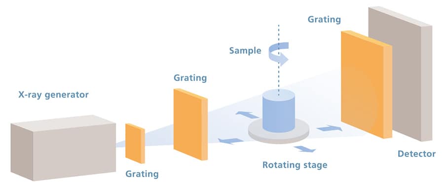
Phase-Contrast X-Ray CT System

The Xctal 5000 is a new X-ray CT system that creates images of X-ray phase shifts.
In addition to the X-ray absorption information detected by conventional X-ray CT systems, this system can detect X-ray scattering and refraction information. This provides observations of fine structure across a wide field of view, and high-contrast observations of samples with no absorption differences.
This is useful for research and development of fiber reinforced resins, composite materials, and biomaterials, which are advancing through research.
A new scanning method has been adopted that can detect phase shifts from X-ray interference using a diffraction grating.
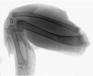
2D projection image (Absorption)
Absorption images visualize X-ray absorption differences, which are also detected by conventional X-ray systems.
This enables detailed observations of shapes within the sample.
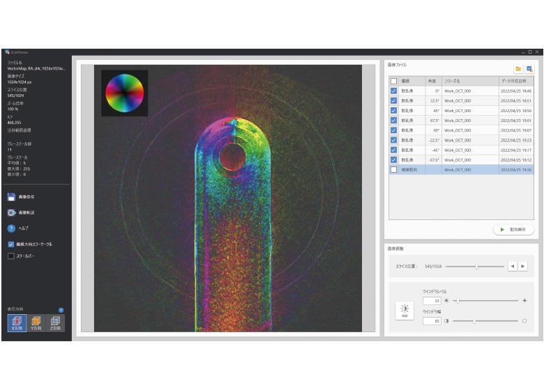
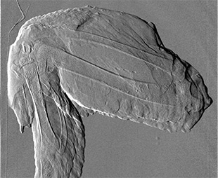
2D projection image (Dark)
Dark-field images visualize dispersion by fine structure.
Fine cracks can be detected with a field of view up to 100 mm in size. In addition, the system is equipped with a fiber orientation analysis function, enabling observations of fiber flow over a wide field of view.
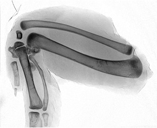
2D projection image (Phase)
Phase images visualize density differences.
The system is capable of high-contrast observations of samples with no absorption differences, including resin products made of different materials.