Benchtop MALDI-TOF Imaging Starter Kit - Applications
Analysis of lipids in full rat brain with 30 µm and 50 µm spacing

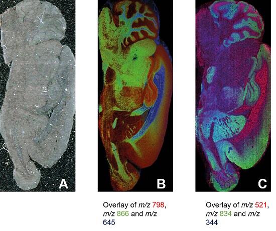
MALDI images of full rat brain using FlexiVision-mini ITO glass slide. A, optical scan; B, positive ion mode image from MALDI-8020 using 50 µm spacing; C, negative ion mode image from MALDI-8030 using 30 µm spacing.
Sample B
| Matrix | DHB, sublimated with Shimadzu iMLayer device |
|---|---|
| Measurement region | 67,024 pixels |
| Measurement time | around 1.9 hours |
| Experiment details | Laser repetition rate of 200 Hz and 20 shots per pixel |
Sample C
| Matrix | 9-AA, sublimated with Shimadzu iMLayer device |
|---|---|
| Measurement region | 275,210 pixels |
| Measurement time | around 3.8 hours |
| Experiment details | Laser repetition rate of 200 Hz and 10 shots per pixel |
Large molecule imaging (protein/ on-tissue digestion) with 50 µm spacing

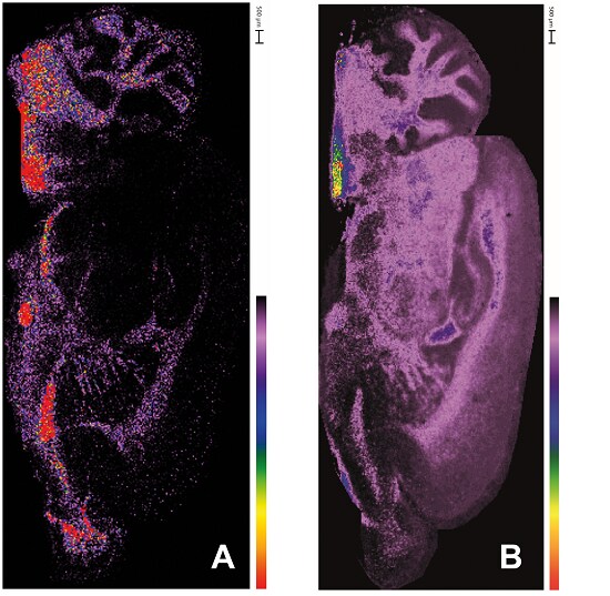
MALDI images of protein and peptides in full rat brain using FlexiVision-mini ITO glass slide and MALDI-8020. A, MBP protein, (m/z 14124); B, MBP peptide (HGFLPR, m/z 726.39)
Sample A
| Matrix | Sinapinic acid, sprayed with Shimadzu iMLayer AERO device |
|---|---|
| Measurement region | 68,836 pixels |
| Measurement time | around 3.8 hours |
| Experiment details | Laser repetition rate of 100 Hz and 20 shots per pixel |
Sample B
| Matrix | CHCA, sprayed with Shimadzu iMLayer AERO device |
|---|---|
| Measurement region | 67,482 pixels |
| Measurement time | around 2.8 hours |
| Experiment details | Laser repetition rate of 200 Hz and 30 shots per pixel |


