Carbon Nanotube | Dispersion | 3D Images of Carbon Nanotubes
A scanning probe microscope (SPM) offers observations of carbon nanotubes (CNTs). It offers comprehensive information, including diameters, lengths, dispersion status and the presence of impurities.
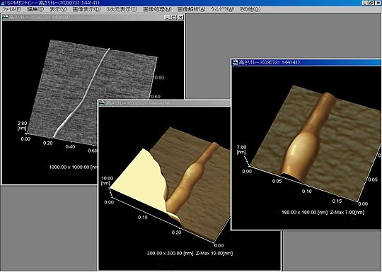
Fig. 1 shows lengths estimated from CNT images.
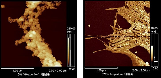
Fig. 2 shows unpurified double-walled carbon nanotubes (DWNTs) immediately after manufacture, and the same DWNTs after purification.
CNTs require purification after manufacture due to the high levels of impurities, such as catalyst and oxide. SPM provides direct observations of CNTs and non-CNT residues before and after purification. It is an effective method for evaluating the purification status. Before purification, the majority of DWNTs cannot be recognized as being CNTs in shape. However, the CNT shapes are apparent for the purified DWNTs in Fig. 2.
The bundling and dispersion can also be observed.
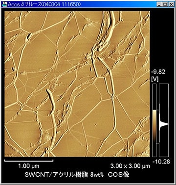
Fig. 3 shows an SPM phase image (COS image).
The elasticity mapping image shows that the SWNT bundles disperse in the resin and spread out to form a network.
(The sample was created by rubbing SWNTs with an imidazolium ionic liquid to form a gel and using a hot press to form a film.)

Fig. 1 Samples Supplied by Central Research Institute of Electric Power Industry

Fig. 2 DWNTs Before and After Purification (Samples supplied by Saito Laboratory, Graduate School of Engineering, Nagoya University)

Fig. 3 SPM Phase Image (COS image) (Samples supplied by Aida Nanospace Project, Japan Science and Technology Agency)
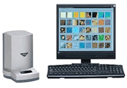
Scanning Probe Microscope

The scanning probe microscope (SPM) scans sample surfaces with a microscopic probe to provide high-magnification 3-dimensional observations.
The SPM-9700 is the next generation of spanning probe microscope that represents further evolution of previous models that were highly regarded for rapid observations and simple operation.


