Observation Examples of Thin Film
Phase image and photo-assisted KFM measurements of zinc oxide: copper phthalocyanine bulk-heterojunction layer prepared by simultaneous vacuum evaporation process
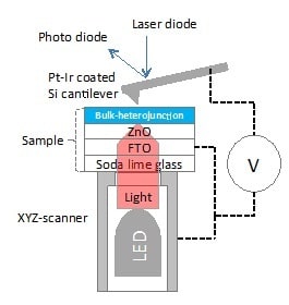
Fig. 1 PKFM Diagram
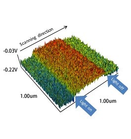
Fig. 2 Potential (3D)
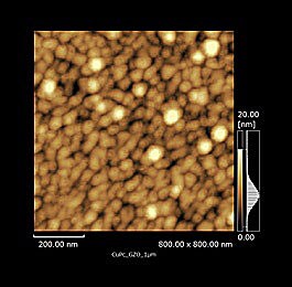
Fig. 3 Height
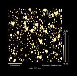
Fig. 4 Phase
The change in the surface potential originated from the increase in free carrier by irradiating light could be measured with a photo-assisted kelvin force microscopy (PKFM) represented by Figure 1 for a Ga-doped zinc oxide: Cu-phthalocyanine bulk-heterojunction layer prepared by a simultaneous vacuum evaporation process, as shown in Figure 2. The distribution of zinc oxide (bright part) and Cu-phthalocyanine (dark part) could be observed for the corresponding topography image (Figure 3) by the phase imaging with a dynamic mode atomic force microscopy, as shown in Figure 4.
M. Izaki, et al., Hybrid zinc oxide: Cu-phthalocyanine bulk-heterojunction photovoltaic device, RSC Advance, 4, 14956 (2014)
(Courtesy of Prof. Masanobu Izaki, National University Corporation, Toyohashi University of Technology Graduate School of Engineering, Faculty of Mechanical Engineering)