Observations of SWNT/Polymer Composite
Surface Observations of SWNT in Polymer
Observations were made of a gel formed into films from a mixture of an imidazolium ionic liquid and single-wall carbon nanotubes (SWNT). The topographic image in Fig.1 shows SWNT unraveling from the bundled state to a web. Fig.2 shows elasticity observations of the same view field. It provides a clearer image of the CNT distribution in the gel.
Surface Potential Measurements of SWNT in Polymer
Fig.3 shows a topographic image of SWNT stretching like a web in the gel. Fig.4 shows the results of simultaneous topographical and surface-potential observations on the same sample, represented as a surface-potential image. The higher potential measured along the SWNT provides clearer observations of the state of the SWNT within the gel.
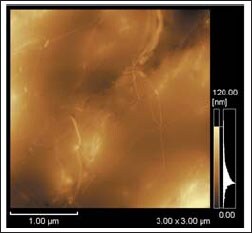
Fig.1 Topographic Image of SWNT in Polymer
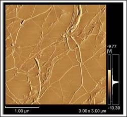
Fig.2 Elasticity Image of SWNT in Polymer
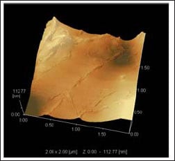
Fig.3 Topographic Image of SWNT in Polymer
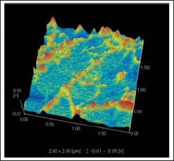
Fig.4 Surface-potential Image of SWNT in Polymer